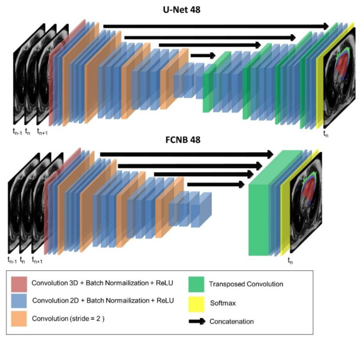
Figure 2.
Networks’ optimized architecture. The two networks evaluated in this study: U-Net and fully-convolutional network (FCNB) architectures included a first 3D convolution layer to allow multiple cardiac frames as input. Following 2D convolution layers encoded images from 48 features up to 768 features. Eventually, the decoder targeted three labels for segmentation in the central input frame: epicardial adipose tissue (EAT), paracardial adipose tissue (PAT), and heart ventricles (HV).

