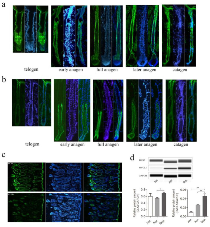
Figure 5.
Detection of spatiotemporal expression of DLX3 and OVOL1 during yak HFs cycle by immunofluorescence. (a,b) The expressions of DLX3 (a) and OVOL1 (b) were detected using anti-Dlx3 and anti-Ovol1 antibody (green), respectively, in hair follicle at telogen, early anagen, full anagen, later anagen, and catagen. Staining in each period was represented by a single hair follicle—blue indicates DAPI staining. (c) Panoramic display of a microscope field in 10× of DLX3 (upper) and OVOL1 (lower) immunofluorescence staining. (d) Western blot analyses for DLX3 and OVOL1 protein levels in Jan. (catagen), Apr. (telogen), and Sep. (anagen), during yak HFs cycle, and the quantitative analysis of gray value were shown in histograms. Data are presented as mean ± SEM for 3 biological replicates; * p < 0.05; ** p < 0.01.

