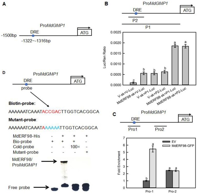
Figure 5.
MdERF98 directly binds to the promoter of MdGMP1. A, Diagram of MdGMP1 promoter region containing a MdERF98 potential DRE binding site, which is located at 1,322- to 1,316-bp upstream of the MdGMP1 initiation codon (ATG). B, Dual luciferase assays in tobacco leaves showed that MdERF98 binds to the promoter of MdGMP1. P1 represents the 1,500-bp promoter of MdGMP1, P2 represents the fragment of the MdGMP1 promoter containing the DRE motif, V-sk represents empty pGreenII 62-SK vector, V-Luc represents empty pGreenII 0800-LUC vector. C, ChIP-qPCR assay in transgenic apple calli. Cross-linked chromatin samples were extracted from 35S:MdERF98-GFP or EV-GFP (Empty pMDC83-GFP vector) transgenic apple calli and precipitated with an anti-GFP antibody. Eluted DNA was subjected to PCR for amplification of sequences neighboring the DRE by qPCR. Pro1 and Pro2, two regions of MdGMP1 promoter, were investigated. The value of Pro-1 EV-GFP was set to 1. The ChIP assay was repeated three times, and the enriched DNA fragments in each ChIP were used as one biological replicate for qPCR. D, EMSA showing that MdERF98 binds to the DRE motif of the MdGMP1 promoter. The biotin-labeled probe was a fragment of the MdGMP1 promoter containing the DRE motif, and the cold-probe was the nonlabeled probe sequence (at 100-fold that of the bio-probe). The mutant-probe was the biotin-labeled sequence with five nucleotides mutated. MdERF98-His was a purified fusion protein. Bars represent the mean value ±se (n = 3). Lowercase letters indicate significantly different values (P < 0.05, one-way ANOVA test).

