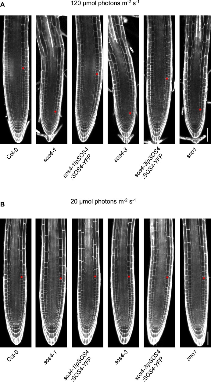
Figure 5.
Root tip morphology of sos4 and wild type under standard and low light conditions. A, PI staining of the root tip zone of 3-d-old seedlings grown on sterile culture medium in 16-h light (120 µmol photons m−2 s−1 at 21°C), 8-h dark (at 18°C) cycles. The images are aligned to the position of the quiescent center. The scale bar represents 50 µm. The red dot marks the first cortical cell of the elongation zone defining the end of the meristem region. B, As in (A) but from seedlings grown under a light intensity of 20 µmol photons m−2 s−1. Each image is representative of n ≥ 20.

