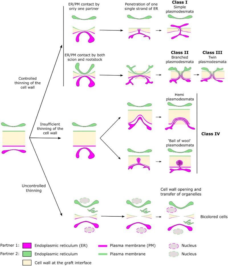
Figure 6.
A model of secondary PD biogenesis at the graft interface. Class I PD could result from unidirectional desmotubule entry by one partner and extreme cell wall thinning. Class II PD could be the consequence of synchronized and symmetric desmotubule entry into a thinned cell wall by both the scion and the rootstock. Subsequently, during cell wall extension, Class II PD could mature into Class III PD. Class IV could correspond to de novo PD formation attempts which are incomplete due to an insufficient thinning of the cell wall at the graft interface. The existence of “ball of wool” shape PD suggests that desmotubule entry into the cell wall is an active mechanism. The presence of PD of Class IV in the same proportions in the scion and the rootstock suggest that both grafting partners are able to initiate PD biogenesis at the graft interface. In addition, bicolored cells at the graft interface could be the consequence of uncontrolled cell wall thinning leading to the formation of cell wall gaps through which nucleus and ER could transit from one partner to the other.

