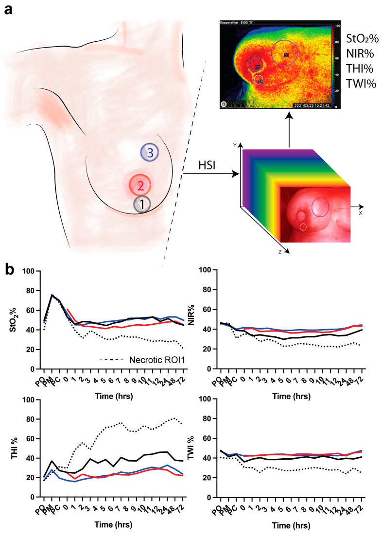
Figure 1.
(a) Regions of interest. ROI-1 (black) is the region of the vertical scar underneath the skin island of the DIEP flap to the inframammary fold; ROI-2 (red) is the skin island of the DIEP flap. ROI-3 (blue) is the medial side of the mastectomy skin flap. Hyperspectral imaging (HSI) is performed to extract the hypercube with which the algorithm extracts 4 parameters (StO2%, NIR%, THI%, and TWI%). (b) HSI acquisition is made pre-operatively (PO), post-mastectomy (PM), post-clip (PC), and every hour for 12 h and every day for 3 days. Data are expressed as mean value.

