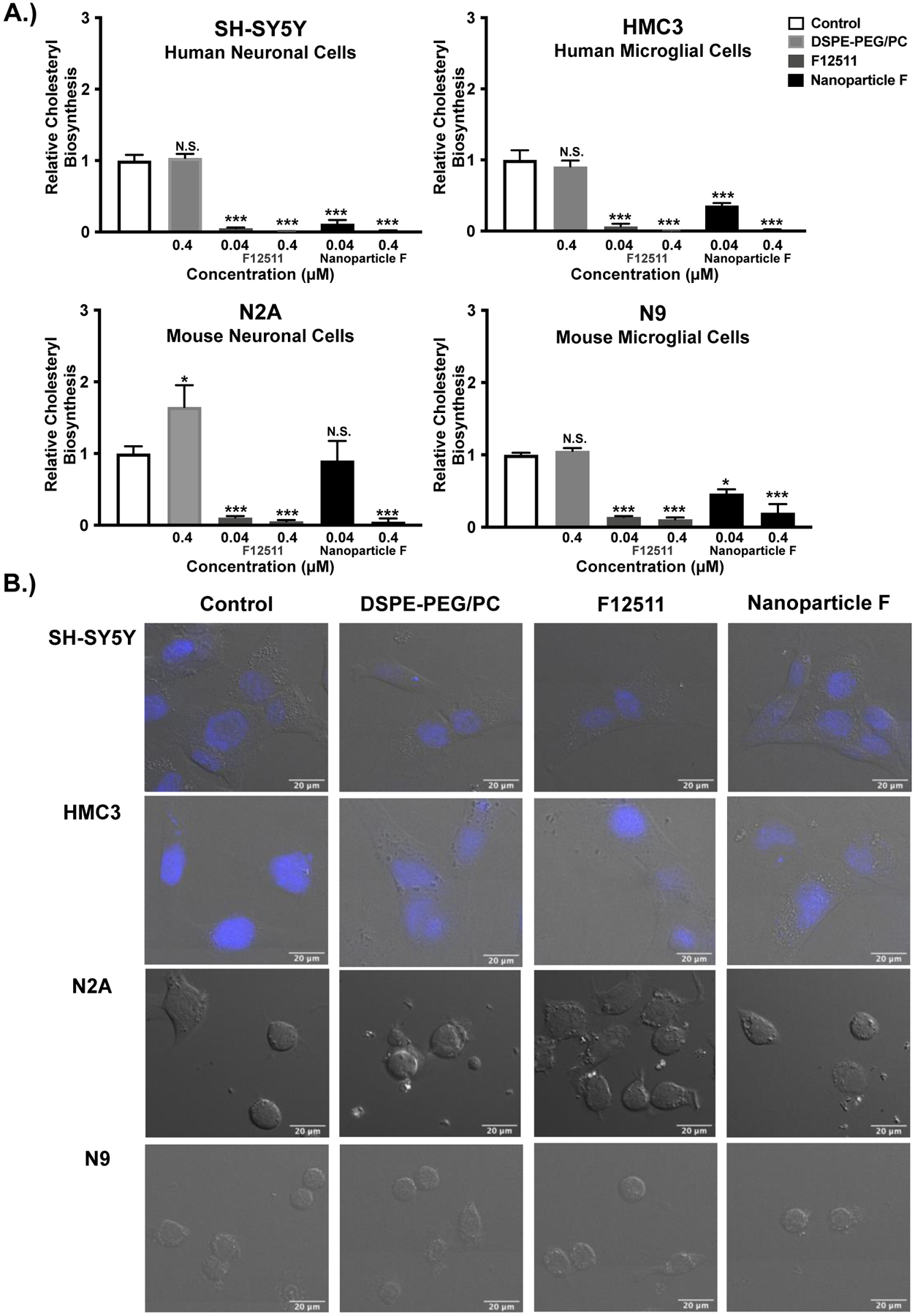
Figure 3:

Nanoparticle F decreases ACAT activity in intact cells of human and mouse origin with no obvious effects on cell morphology. Results and images from human neuronal (SH-SY5Y), human microglial (HMC3), mouse neuronal (N2a), and mouse microglial (N9) cells. A.) Cells at 80–90% confluency in triplicate dishes (12- or 6-well dishes) were treated for 2 hours with EtOH (control) or with the following agents: 0.4μM DSPE-PEG2000/PC; 0.04μM or 0.4μM of F12511 alone or of Nanoparticle F. The cells were then pulsed with 3H-oleate to determine ACAT activity as described in the Materials and Methods section. One-way ANOVA statistical test was completed, and results shown are relative to the control. N.S. not significant; p<0.001 ***, p<0.01 **, p<0.05 *. B.) Cells as indicated were treated for 2 hours with 0.5μM EtOH (control), DSPE-PEG2000/PC, F12511 alone (EtOH) or Nanoparticle F and imaged. Representative images of DIC and Hoechst (blue, top 2 rows). Images were analyzed using ImageJ. Scale bar is 20μm.
