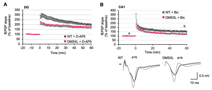
Figure 6.
Contribution of NMDA and GABA receptors to altered LTP in DMSXL mice. (A) LTP in DG in the presence of the NMDA antagonist D-AP5 (0.5 µM) (DMSXL, n = 8 mice, n = 18 slices; WT, n = 7 mice, n= 16 slices). APV was added to the superfusion bath 20 min before high-frequency stimulation and during the entire recording. D-AP5 did not restore the lower strength of LTP in DMSXL mice. (B) LTP in the CA1 area in the presence of the GABAA antagonist Bic (4 µM) in DMSXL (n = 4 mice, n = 9 slices) and WT (n = 3 mice, n = 7 slices) mice. Bic was added to the perfusion bath 20 min before high-frequency stimulation and during all the recording. Bic did not restore the lower strength of LTP in DMSXL mice. Representative superimposed sample traces of evoked AMPAR-mediated fEPSPs before (a) and after (b) completion of LTP in a slice from WT and a slice from DMSXL mice.

