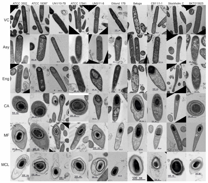
Figure 1.
Electron micrographs of sporulating Clostridium botulinum cells. Images of thin sections of fixed, sporulating C. botulinum cultures were acquired to observe the ultrastructure of sporulating cells. The cells were categorized according to their morphological stages: VC, vegetative cell; Asy, asymmetric division; Eng, engulfment; CA, coat assembly; MF, mature forespore; MCL, mother cell lysis. Exosporia visible while the forespore was still in the mother cell are indicated with black arrows. Scale bars represent 500 nm. Enlarged versions of these images can be found in Figures S2 and S3.

