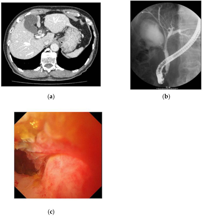
Figure 1.
Images of the case with bile wall thickness which was finally diagnosed as IgG4SC: (a) The CT scan revealed wall thickening in the common bile duct, with nodular thickening of the bile ducts in the hilar region; (b) the ERCP showed that the wall of the bile duct was hard, and the mucosal edge was rough from the cystic duct to the hilar region of the bile duct; (c) cholangioscopy showed rough mucosa with irregular papillary elevation.

