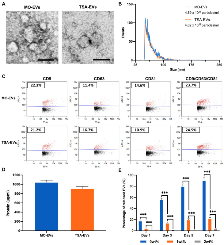
Figure 4.
Characterisation of EVs derived from TSA treated and untreated mineralising osteoblasts. (A) TEM image of isolated EVs. Scale bar = 50 nm. (B) Nano-flow cytometry (NanoFCM) analysis, depicting the size distribution and concentration of particles. (C) Single-particle phenotyping of osteoblast-derived EVs. EVs were fluorescently labelled with APC-conjugated antibodies specific to CD9, CD63 and CD81. Bivariate dot-plots of indicated fluorescence versus SSC are shown. In addition, CD9/CD63/CD81 positive particles are shown. (D) EV protein content. (E) Quantification of EVs released from GelMA hydrogel with/with LAP assessed via CD63 positive ELISA. Data are expressed as mean ± SD (n = 3). *** p ≤ 0.001.

