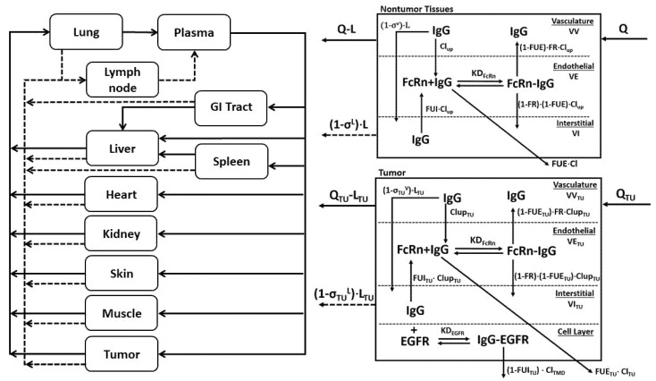
Figure 7.
Schematic representation of the PBPK Model: The PBPK model is shown as a schematic diagram with solid lines representing plasma flow to and from tissues and dashed gray lines representing lymph flow. B. Each tissue compartment is composed of three sub-compartments representing vascular, endothelial, and interstitial spaces. Q and L represent tissue plasma and lymph flow, and σV and σL represent the vasculature and lymph reflection coefficients. Endosomal uptake is represented as Clup, and endosomal recycling is the product of Clup and the fraction of FcRn bound mAb that is returned to the vasculature, abbreviated as FR. KDFcRn is the equilibrium dissociation constant for FcRn-mAb binding. Organ-specific elimination of unbound mAb in the endothelial space (FUE) is denoted as Cl. C. The tumor sub-compartment structure is shown. A 4th layer representing the cellular fraction is added, allowing the antibody to bind its target antigen (i.e., EGFR) with the observed equilibrium dissociation constant (KDEGFR) with bound antigen eliminated through receptor internalization and degradation (ClTMD).

