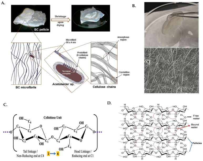
Figure 1.
(A) Arrangement of microfibril in amorphous and crystalline region and its macroscopic appearance in wet conditions. (B) BC loaded with water and its SEM image. (C) Molecular structure of “cellobiose unit.” (D) H-bonding in the matrix of the BC. Reproduced with permission from [27] (Copyright © 2022, Elsevier), [32] (Open Access).

