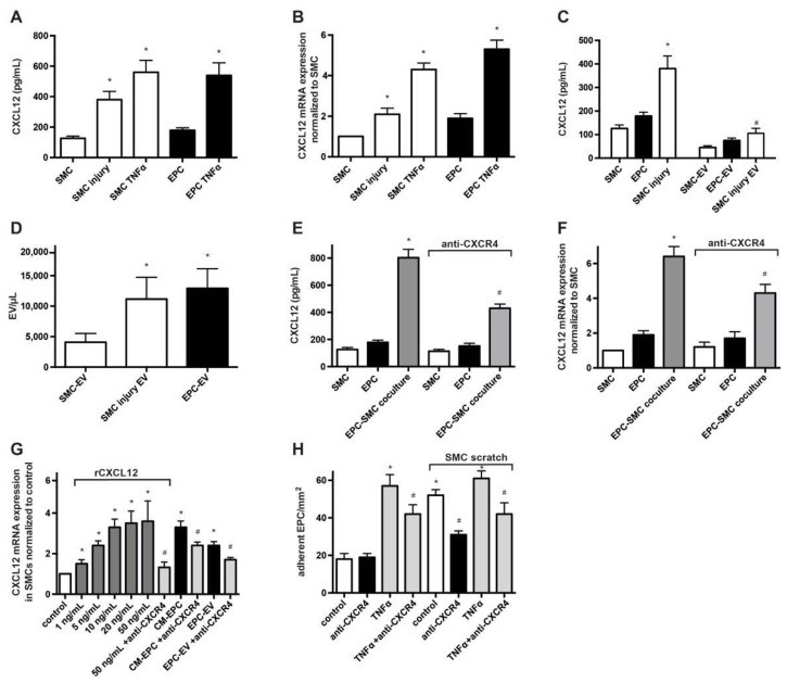
Figure 1.
Expression and release of CXCL12 by SMCs and EPCs. (A) Analysis of CXLC12 release using ELISA. Cell supernatants from monocultured SMCs and EPCs, treated as indicated, were collected 24 h after cultivation. * p < 0.05 vs. untreated cells; n = 6. (B) Real-time RT-PCR analysis of CXCL12 expression SMCs and EPCs treated as indicated. Results were normalized to CXCL12 expression in SMCs. * p < 0.05 vs. untreated cells; n = 5. (C) Detection of CXCL12 in MVs derived from monocultured SMCs and EPCs. Isolated MVs were lysed in RIPA buffer and CXCL12 levels were determined using ELISA. * p < 0.05 vs. non-injured SMCs, # p < 0.05 vs. SMC-MV; n = 5. (D) Enumeration of MVs in the supernatant of EPCs, non-injured SMCs and injured SMCs using flow cytometry with calibrated microbeads. * p < 0.05 vs. non-injured SMCs; n = 4. (E) Evaluation of the effect of EPC-SMC co-cultivation and engagement of CXCR4 on the release of CXCL12. Supernatants from monocultured SMCs, monocultured EPCs and EPCs co-cultured with SMCs, each in the presence or absence of a blocking CXCR4 Ab, were analyzed for CXCL12 concentration using ELISA. * p < 0.05 vs. SMCs, # p < 0.05 vs. EPC-SMC co-culture in the absence of anti-CXCR4; n = 5. (F) Real-time RT-PCR analysis of CXCL12 expression to test the impact of EPC-SMC co-cultivation and involvement of CXCR4. CXCL12 transcripts were determined in SMCs, EPCs and EPC-SMC co-cultures in the presence or absence of a blocking CXCR4 Ab. * p < 0.05 vs. SMCs, # p < 0.05 vs. EPC-SMC co-culture in the absence of anti-CXCR4; n = 5. (G) Real-time RT-PCR analysis of CXCL12 expression in SMCs treated with various doses of rCXCL12, CM-EPC or EPC-MV in the presence or absence of an anti-CXCR4 Ab. * p < 0.05 vs. untreated SMCs (control), # p < 0.05 vs. respective treatment in the absence of anti-CXCR4; n = 5. (H) Adhesion of EPCs to SMCs under flow conditions in vitro. EPCs pretreated with/without an anti-CXCR4 Ab were perfused in a parallel flow chamber and the number of cells EPCs adherent to the SMC monolayer was determined and expressed as adherent cells per 1 mm². For some experiments, the SMC monolayer was wounded by a linear scratch before perfusion of EPCs. * p < 0.05 vs. untreated and non-scratched SMCs (control), # p < 0.05 vs. respective treatment in the absence of anti-CXCR4; n = 4 to 6.

