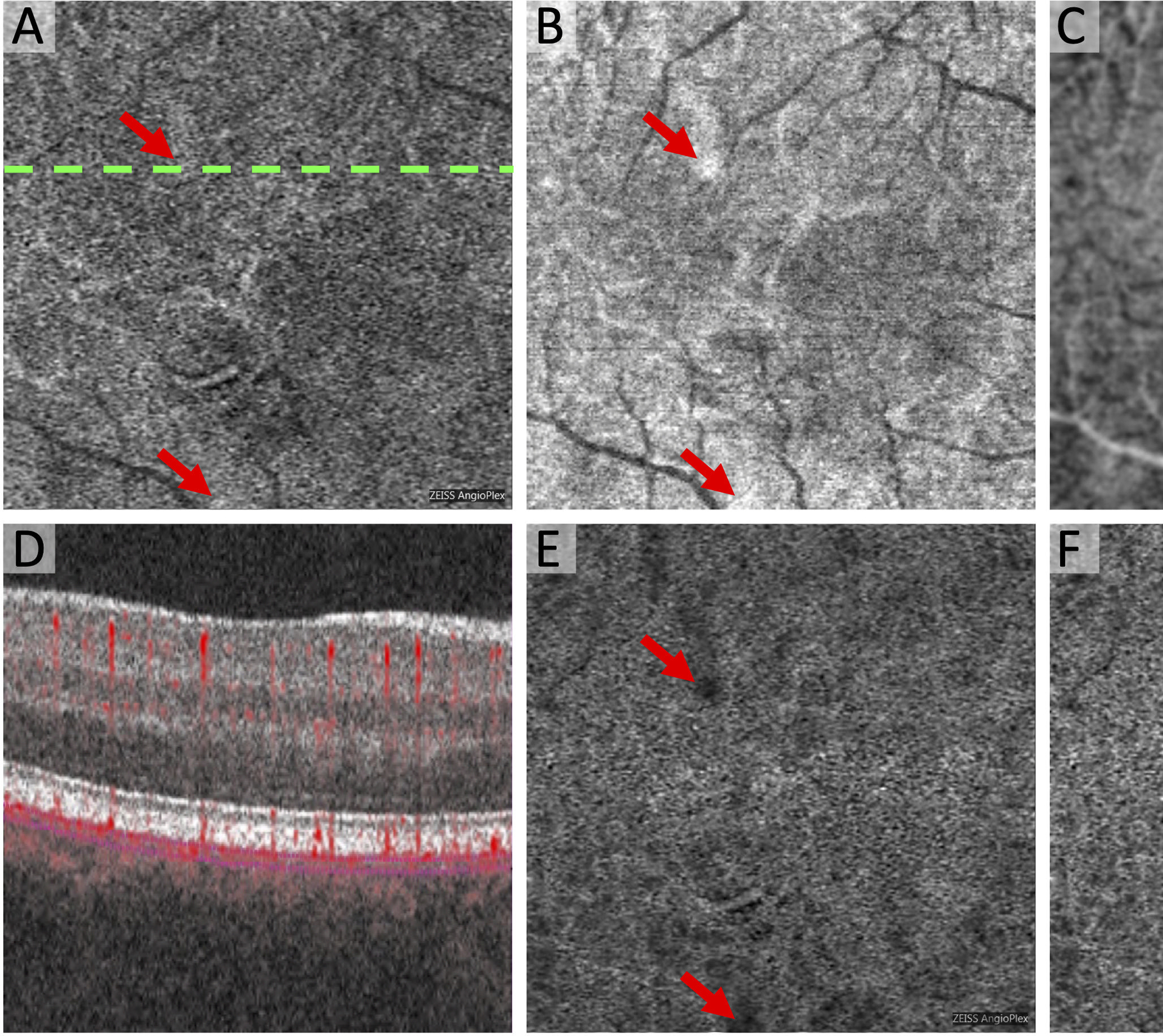
Figure 2. Compensation for Signal Attenuation Introduces Artifactual Flow Deficits in the Choriocapillaris.

(A) Original Zeiss Angioplex OCTA of the choriocapillaris with dotted line showing location of OCTA B-scan in (D). (B) Original structural OCT of the choriocapillaris with same segmentation as (A) and areas of hyperreflectivity (arrows). (C) Inverted and blurred structural OCT now with areas of relative hyporeflectivity (arrows). (E) Compensated OCTA as a result of multiplication of (A) x (C). (F) Final OCTA image with global compensation for Q-score showing newly introduced flow deficits that are not present in the original OCTA (A). These artifacts are related to the hyporeflective areas in the inverted structural OCT (arrows in C).
