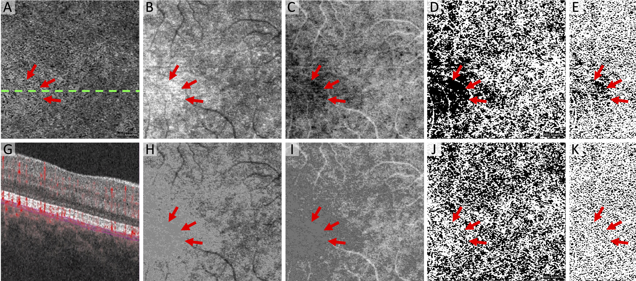
Figure 4. Modified Approach Reduces of Artifactual Flow Deficits Caused by Image Compensation.

(A) Original Zeiss Angioplex choriocapillaris OCTA flow image with dotted line indicating location of cross-section below showing segmentation lines (G). (B and H) Choriocapillaris structural OCT images. (C and I) Inverse of the structural OCT used to compensate for signal attenuation. Note inverted slab is Gaussian blurred before multiplication with OCTA. (D and J) MinError(I), (E and K) Phansalkar, and (F and L) Li thresholded images after compensation. Bottom row uses the modified algorithm for eliminating hyporeflective regions in the inverse OCT and the top row does not, which leads to large new flow deficits (arrows).
