
Fig. 4. Decreased concentrations of ER membrane cholesterol in human VSMCs Wnt5a -/−.
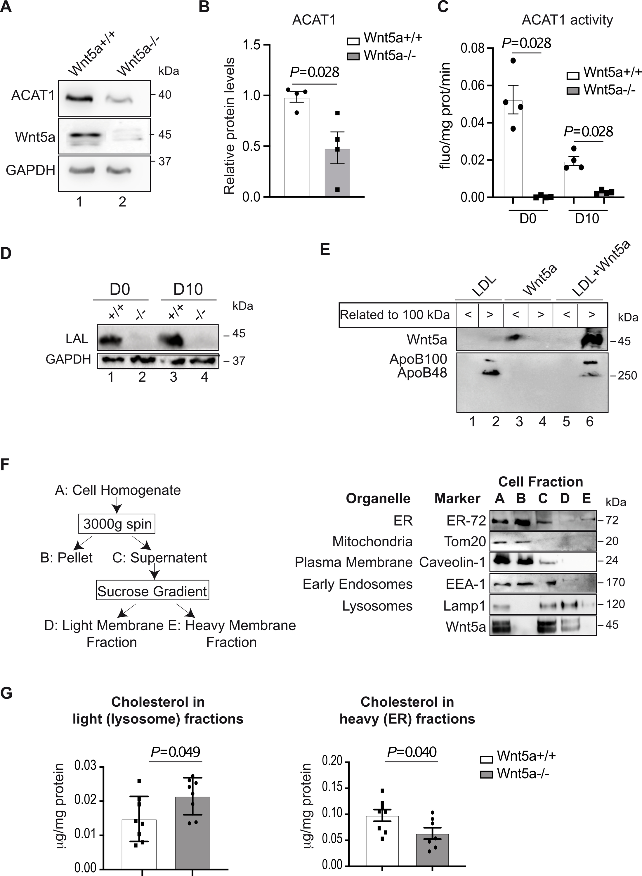
A, A representative immunoblot shows the protein expression of ACAT1 and B, its quantification in human Wnt5a−/− VSMCs vs controls upon cholesterol accumulation protocol calculated as relative to GAPDH expression (n=3). C, ACAT1 activity in human Wnt5a−/− VSMCs vs controls before (D0) and upon (D10) cholesterol accumulation treatments during 10 days (n=4). D, A representative immunoblot shows the protein expression of the Lysosomal acid lipase (LAL) (n=4). E, Wnt5a was incubated with human LDL particles as described in the method section. A representative immunoblot shows the presence of Wnt5a in the <100 kDa fraction when incubated with CHAPS, or in >100 kDa apoB positive fraction when incubated with human LDL (n=3). F, Purification of ER and LELs, purification of mitochondria, plasma and early endosomal membranes. Left panel is showing the diagram of LELs membrane fractionation scheme. A-E denote major fractions recovered and analyzed by western blot. Right panels, VSMCs were treated according to the fractionation scheme and as described in the method section. Aliquots representing equal volumes of each fraction (A-E) were subjected to immunoblot analysis for the indicated organelles markers and Wnt5a. G, Quantification of cholesterol concentrations in heavy (E, ER) and light (D, lysosomes) membrane fractions from human Wnt5a−/− VSMCs and Wnt5a+/+ (n=8 and n=7 Wnt5a−/− heavy fractions). B, C and G show individual data along with mean±SEM. For B, C and G, data were analyzed using a Shapiro-Wilk test to assess normality followed by Mann-Whitney test. For B, P=0.028; C, P=0.028 and G, P=0.049 and P=0.040 indicate significance relative to Wnt5a+/+.