
Fig. 5. Wnt5a decreases mTORC1 activity.
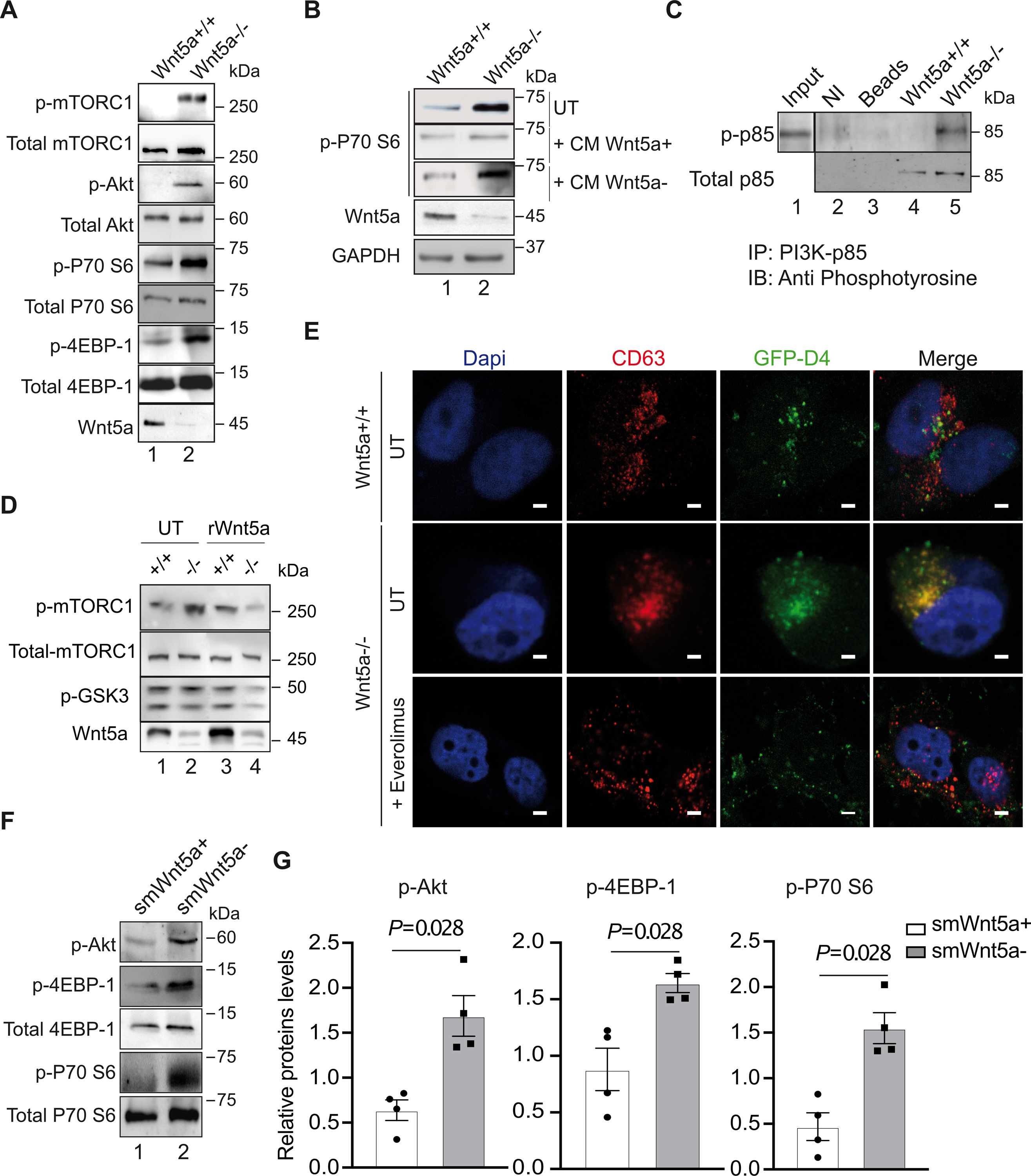
A, shows a representative immunoblot of the indicated genes in human Wnt5a−/− and control VSMCs upon cholesterol accumulation (n=3). B, Western blot analysis showing p-P70 S6 (Thr389) expressions in Wnt5a−/− VSMCs untreated (UT) or treated with conditioned medium enriched in Wnt5a (CM Wnt5a+) or mock medium (CM Wnt5a−) as described in the method section (n=3). C, Immunoprecipitation in Wnt5a−/− VSMCs and controls of the PI3K-p85 subunit followed with immunoblotting with anti-phosphotyrosine (4G10) antibodies show the tyrosine-phosphorylated form of p85 (p-p85) in the absence of Wnt5a. NI=non-immune antibodies, Beads = empty beads. Input is from whole cell lysate (n=3). D, A representative immunoblot shows p-mTORC1, total mTORC1, p-GSK3, and Wnt5a expressions in Wnt5a−/− VSMCs (−/−) and control cells (+/+) untreated (UT) or treated (rWnt5a) with human recombinant Wnt5a (n=3). E, Confocal analysis showing colocalization (yellow) between GFP-D4 positive vesicle (cholesterol, green) and CD63 (LELs, red) in untreated (UT) and Everolimus® (30nM) treated Wnt5a−/− VSMCs. Scale bars are 5μm (n=3). F, Western blot analysis and G, quantification of relative protein levels of the indicated genes in aortas from control (smWnt5a+) and mutant (smWnt5a−) mice (n=4 mice/group). G shows individual data along with mean±SEM. Data were analyzed using Shapiro-wilk followed by Mann-Whitney test. G, P=0.028 indicate significance relative to smWnt5a+ mice.