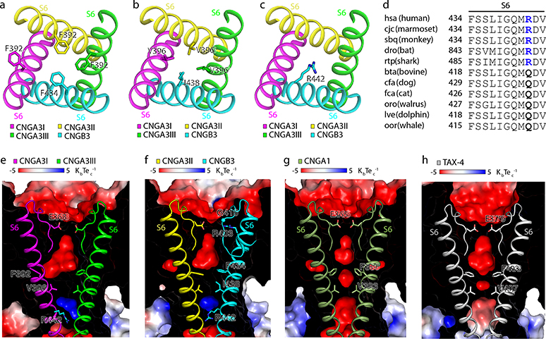
Fig. 4 |. The cavity gate and inner gate.
a, b, Cavity gate in S6 (viewed from the extracellular side). c, Inner gate in S6 (viewed from the extracellular side). d, Amino acid sequence alignment of the gate-forming region of S6 of B3 from different species. Sequences and species abbreviations were obtained from the KEGG database (http://www.genome.ad.jp/kegg/). e-h, Electrostatic potential (calculated with APBS plug-in in PyMol) of the interior surface of the ion conduction pathway in apo A3/B3 (e, f), CNGA1 (g, PDB ID: 7LFT), and TAX-4 (h). Front halves of the channels were cut away and rear halves were overlayed with the SF and S6.

