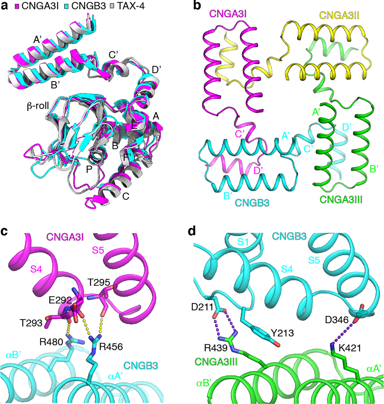
Fig. 6 |. The C-linker and CNBD.

a, Comparison of the structures of the C-linker and CNBD in apo A3I, B3 and TAX-4. b, The gating ring viewed from the extracellular side. c, Interactions between helices A’B’ of the C-linker of B3 and S4-S5 of A3I. Hydrogen bonds are shown as yellow dash lines. d, Interactions between helices A’B’ of the C-linker of A3III and the TMD of B3. Salt bridges are shown as purple dash lines.
