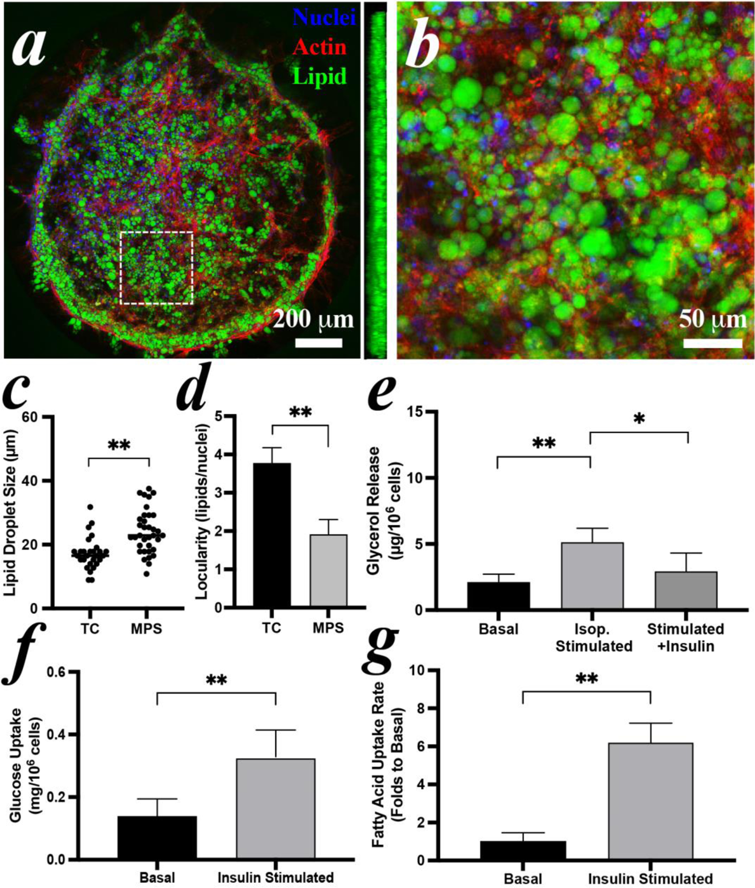
Figure 6: Reconstitution of hMSCs-derived iADIPO-MPS.

(a) morphology of adipocytes in MPS and (b) zoomed view of white dash-lined area in (a). A sideview of 3D reconstruction in (a) is inserted aside. (c) lipid droplet size and (d) locularity of the adipocytes differentiated in TC condition and in MPS. Average diameters are 16.5 μm in TC and 22.9 μm in MPS. (e) lipolysis and (f) glucose uptake of the adipocytes in the MPS. (g) analysis of fluorescent fatty acid uptake by adipocytes in MPS with or without insulin stimulation. Pre-induced hMSCs were loaded in MPS at Day 4 and imaged or assayed at Day 14.
