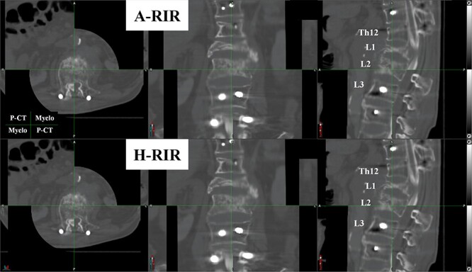
Fig. 2.

An example of the blind review comparing two registration results. Each patient’s registrations were displayed on the horizontal split screen with automatically shuffled. Three evaluators are prompted to choose the registration they would like to use clinically. The figure is shown on a checkerboard display (the planning CT: the upper left and lower right parts, the CT-myelogram: the upper right and lower left). The evaluators can review all the slices of CT images. A-RIR: automated rigid image registration, H-RIR: human rigid image registration, P-CT: planning CT, Myelo: CT-myelogram.
