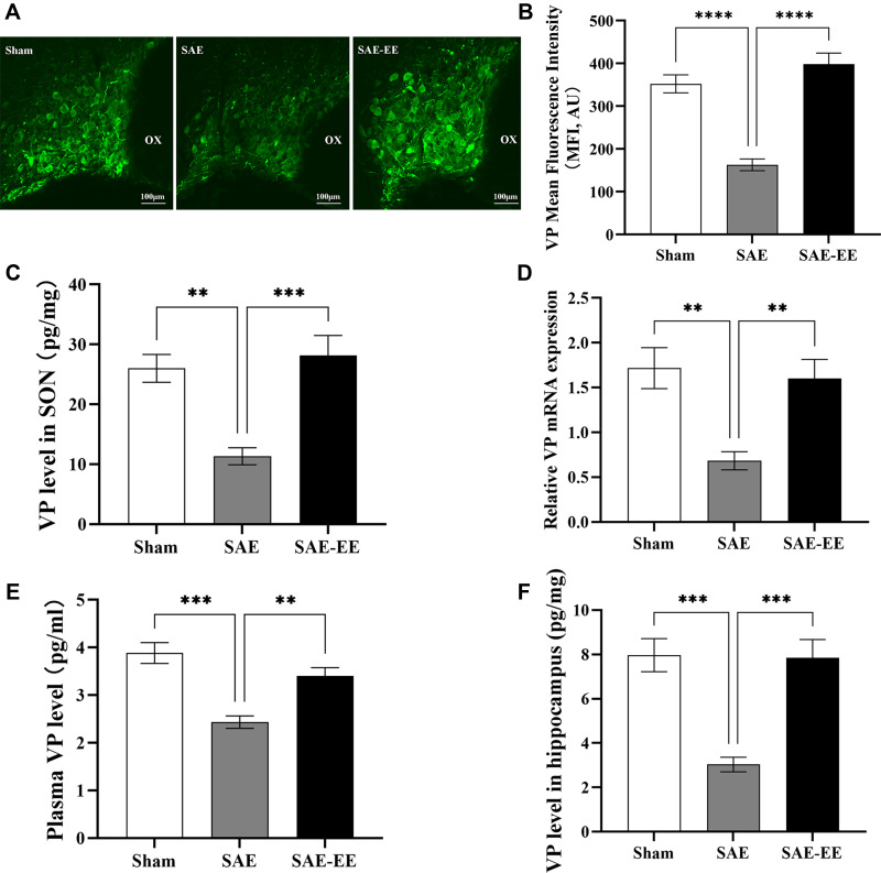
Figure 3.
(A) VP staining in the SON. (B) The VP mean fluorescence intensity in the SAE rats was significantly lower than that in the sham or SAE-EE rats. (C) The amount of VP in the SON in the SAE rats was lower than that in the sham or SAE-EE rats. (D) The level of Vp mRNA in the SON was lower in the SAE rats compared with that in the sham or SAE-EE rats. (E) The plasma VP concentration in the SAE rats was lower than that in the sham or SAE-EE rats. (F) The amount of VP in the hippocampus of SAE rats was lower than that of the sham or SAE-EE rats. **p < 0.01, ***p < 0.001, ****p < 0.0001. Data represent the means ± standard error of the mean.
Abbreviations: VP, vasopressin; SON, supraoptic nucleus; SAE, sepsis-associated encephalopathy; EE, environmental enrichment; Magnification, 40×.

