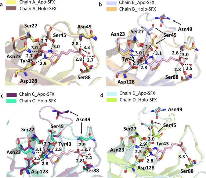
Fig. 3. Superposition of the biotin-binding sites for each chain of the Apo-SFX and Holo-SFX structures.
Chains A–D of Apo-SFX is superposed with the Holo-SFX (PDB ID:5JD2) structure of streptavidin in a–d, respectively (Supplementary Table 1). The L3/4 opening as a “lid” without selenobiotin binding. Binding of selenobiotin is not symmetric for all four monomers, which represent cooperativity. Selenobiotin and water molecules were represented by light pink sticks and red-colored spheres, respectively. Hydrogen bonds are shown with black dashed lines and their corresponding distance as a unit of Angstrom (Å).

