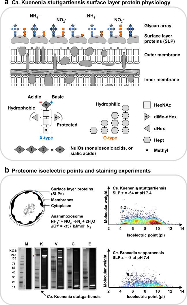
Fig. 5. Physiology of the Ca. Kuenenia stuttgartiensis surface layer protein (SLP) and oligosaccharides.
a The surface layer protein (SLP) of Ca. Kuenenia stuttgartiensis is densely covered by two entirely different types of oligosaccharides (“X-type” and “O-type”, SI-DOC-Fig-S16). The dense layer supposedly provides shielding of the very acidic SLP. Interestingly, the investigated Ca. Kuenenia stuttgartiensis strain produces nonulosonic acids (NulOs), which possess an unmasked amine. Those have the potential to counterbalance the carboxylic acid groups. The sugar symbols are depicted in generic white and gray shades because a further classification into specific types of monosaccharides, beyond sum formulae, chain length and modifications, cannot be obtained from accurate mass experiments. The different shades of gray were simply chosen to make the individual sugars more distinguishable within the oligosaccharide structures. The oligosaccharide structures depicted on the cell surface layer (top graph) are colored in blue if those structures represent a X-type (complex type) structure, or in orange if they represent an O-type (oligo-heptosidic) oligosaccharide. The colors do not provide any further indications on the types of monosaccharides. b The Ca. Kuenenia stuttgartiensis surface layer protein shows a predicted pI (isoelectric point) of ~4.25 and a net charge of ~−60 at physiological pH. In fact, the surface layer protein is one of the most acidic proteins of the complete Ca. Kuenenia stuttgartiensis proteome. On the other hand, the putative surface layer protein of Ca. Brocadia sapporoensis has a predicted pI of only 5.4 and a substantially lower net charge of ~−8 at physiological pH. Moreover, Ca. Brocadia, uses also only a related form of the complex-type oligosaccharide to cover its much less acidic surface layer protein. The SDS-PAGE analyzes show protein and sugar staining for the protein extracts from Ca. Kuenenia stuttgartiensis and the additional control strains H. volcanii (glycan-positive control), C. jejuni (glycan-positive control) and E. coli K12 (glycan-negative control). The left lanes each show the total protein staining (P; Brilliant Blue G staining solution), whereas the right lanes each show the carbohydrate staining (C; Pro-Q 488 Emerald staining kit) “(color figure online)”.

