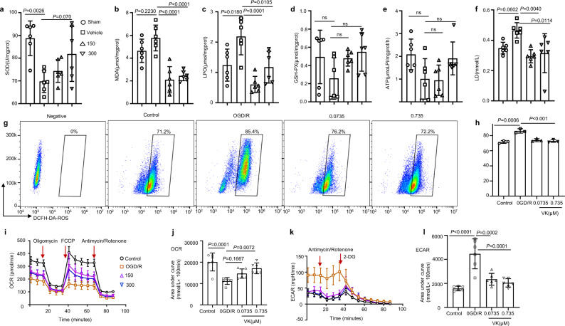
Fig. 3. Effects of VK on oxidative stress and energy metabolism in stroke mice.
a Experimental design: in addition to the sham group, mice (8–10 weeks, male) were subjected to MCAO for 60 min. Mice were randomized into four groups: sham group, vehicle group, and VK (150 and 300 µg/kg) groups. Mice were administered VK at 0, 4, 22.5, and 46.5 h after MCAO/R as assessed by Kits. After reperfusion for 48 h, ischemic tissue was collected. b–d The superoxide dismutase (SOD) activity and malondialdehyde (MDA), lipid peroxide (LPO), and glutathione (GSH-PX) levels in the brain were demonstrated by the use of biochemical kits. Adenosine triphosphate (ATP synthase) (e) and lactic acid (LD) (f) were detected to examine the effect of VK treatment on energy metabolism after reperfusion injury. g HT22 cells stained with DCFH-DA for flow cytometry, using without DCFH-DA as a negative control for the FACS gating strategy. h Quantification of DCFH-DA–positive cells as the mean ± SEM from three independent experiments. DCFH-DA (10 μM). i, j Real-time changes in the O2 consumption rate of neurons in response to treatment with the indicated concentrations of VK for 24 h. Cells were treated with 2 μM of oligomycin, 5 μM of carbonyl cyanide-ptrifluoromethoxyphenylhydrazone (FCCP), and 1 μM of rotenone and antimycin, as indicated by the three red arrows. k, l To assess the extracellular acidification rate, cells were treated with 1 μM of rotenone and antimycin and 50 mM of 2-deoxy-d-glucose (2-DG) as indicated by the two red arrows. Statistical analyses were performed using the Kruskal–Wallis test with the Dunn post hoc test or two-way repeated-measures ANOVA followed by Bonferroni’s post hoc test. Data (animal experiment) represent the mean ± SD, n = 5–7. .

