Figure 6.
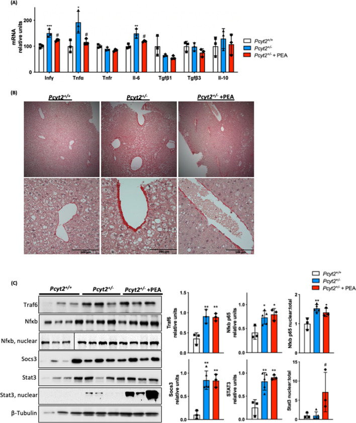
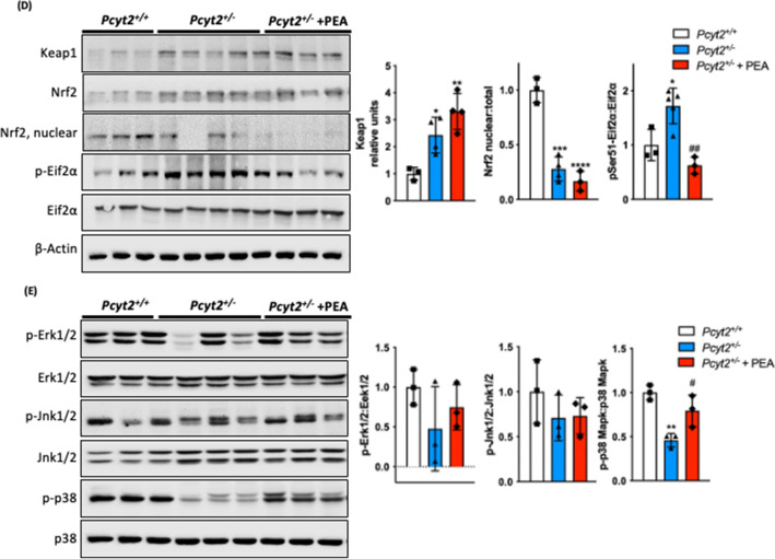
PEA reduces hepatic inflammation. (A) mRNA expression of liver pro- and anti-inflammatory genes in 8-mo Pcyt2+/+, Pcyt2+/− and Pcyt2+/− + PEA mice treated for 12 weeks (n = 3). GAPDH used as loading control. (B) Picrosirius stain of showing that 8-mo Pcyt2+/− liver have increased collagen deposition that could be reversed with PEA. (C) Immunoblot analysis of transcription factors from Jak/Stat and Nfκb pathways: Traf6, Nfκb-p65, Socs3, Stat3; (D) Keap1/Nrf2 and pEif2α activation: Keap1, Nrf2, pSer51-Eif2α, Eif2α and (E) Stress kinases: p-Erk1/2, Erk1/2, p-Jnk1/2, Jnk1/2, p-p38 Mapk, p38 Mapk in 8-mo Pcyt2+/+, Pcyt2+/− and Pcyt2+/− + PEA mice (n = 3–4). Nfκb-p65 nuclear Pcyt2+/+ bands are from a gel separate from the Pcyt2+/− and Pcyt2+/− + PEA bands, but were ran under the same conditions. Band intensities were measured using ImageJ. Membranes were cut prior to antibody hybridization to allow for the probing of multiple targets on one membrane. Data are presented as mean ± SD. *p < 0.05, **p < 0.01, ***p < 0.001 relative to Pcyt2+/+; #p < 0.05, ##p < 0.01 relative to Pcyt2+/−.


