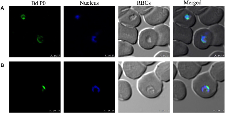
Figure 3.
Cellular localization of BdP0 in the erythrocytic stage. Bdp0 in thin blood smears of B. divergens-parasitic RBCs stained with Bdp0-immune sera under confocal laser microscopy. Anti-rBdp0 serum reactivity with single intracellular parasite forms (A) and sequentially dividing forms (B). Pre-immune and anti-GST sera were used as negative control sera to validate the test (data not shown). Scale bar: 7.5 μm.

