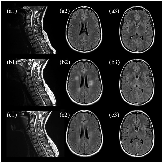
Figure 1.
MRI upon admission (a), early follow-up 5 days after admission (b), and follow-up at 3 months (c). Initial sagittal T2-weighted spinal images with hyperintense lesions extending from C6 to T1 as well as T3 and T4 (a1) and no abnormalities on axial fluid-attenuated inversion recovery images of the brain (a2, a3). MRI at 5-day follow-up showing progressive spinal lesions with additional involvement of c3 to c5 (b1) and new hyperintense lesions of the subcortical white matter (b2) and bilateral pulvinar (b3). Partial resolution of former findings in cervical spine (c1), subcortical white matter (c2), and bilateral pulvinar (c3) at 3 months.

