FIGURE 7.

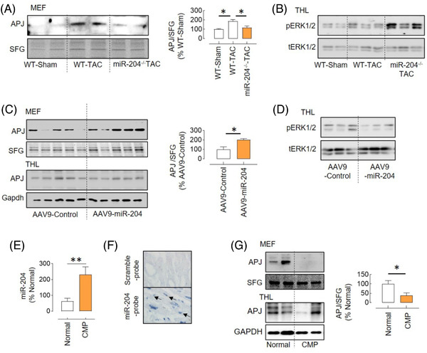
miR‐204 regulation of APJ trafficking in vivo. (A) The APJ expression in the MEF of WT‐TAC and miR‐204–/– TAC mice and its quantification (n = 4). (B) Immunoblot showing increased levels of pERK1/2 in the heart of miR‐204−/− TAC mice compared to WT‐TAC mice. (C) The APJ expression in the MEF of the heart of miR‐204–/– TAC mice receiving AAV9‐miR‐204 or AAV9‐control virus and its quantification (n = 5). (D) Immunoblot showing decreased levels of pERK1/2 in the heart of miR‐204–/– TAC mice provided with AAV9‐miR‐204 compared to those that received control virus. (E) miR‐204 expression in the heart of cardiomyopathy (CMP) patients or normal donors (n = 9–10). (F) In situ hybridisation showing miR‐204 (arrows) in cardiomyocytes of the cardiomyopathy patient heart. (G) The APJ expression in the MEF and total heart‐tissue lysate (THL) of normal donors and CMP patients and its quantification (n = 5). *p < .05, **p < .01 versus indicated group. Data are shown as mean, and error bars represent SEM. SFG, stain‐free gel; scramble‐probe, scrambled control probe
