
Fig. 1. Experimental methods.
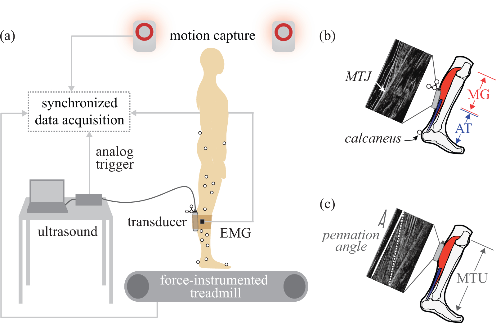
(a) B-mode ultrasound was collected synchronously with motion capture, electromyography (EMG), and ground reaction force data. (b) MTJ tracking. The ultrasound transducer (gray box) was placed over the MTJ of the MG muscle and AT. AT length change was estimated as changes in the straight-line distance from the MTJ to the calcaneus. (c) MF tracking. The transducer was placed over the MG muscle belly. Longitudinal MG length change was calculated from MF length (dotted white line) corrected by the cosine of the pennation angle. Pennation angle was defined as the angle between MF and the superficial fascia (solid white line). MTU length change was estimated from a regression equation based on joint angles.