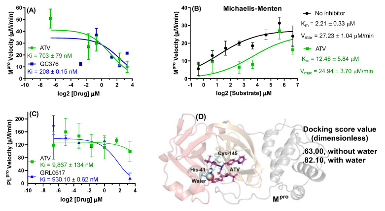
Figure 2.
(A) The ATV and GC376 (positive control) activity on 88.8 nM Mpro velocity at 0–10 μM of inhibitor. (B) Michaelis–Menten plot for 88.8 nM Mpro incubated with substrate concentrations from 0 to 100 μM in the presence and absence of 2.5 μM of ATV. (C) The ATV and GRL0617 (positive control) activity on 100 nM PLpro velocity at 0–10 μM of inhibitor. (D) The 3D representation of the best docking pose for ATV into Mpro catalytic site in the presence of the catalytic water (H2Ocat). For better interpretation the Mpro structure was represented only in the monomeric form with the domains I, II and III in light red, orange, and gray, respectively.

