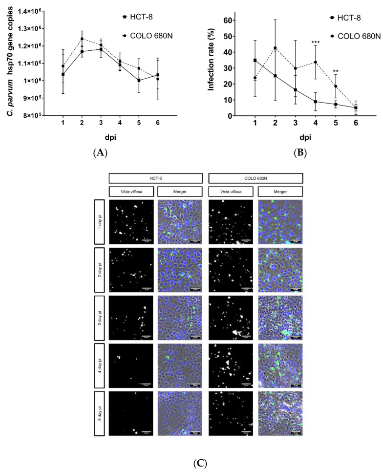
Figure 2.
Replication of C. parvum in HCT-8- and COLO-680N cells using infection protocol III. Parasite intracellular replication was quantified by both qPCR- (A) and Vicia villosa (VV) lectin (VVL)-based immunofluorescence analyses (B). (C) Exemplary illustration of both in vitro cell culture systems: fluorescence-based detection of C. parvum via VVL (green) and cell nuclei via DAPI (blue). To assess parasite development, a two-tailed t-test was performed, comparing the infection rate per day, measured in HCT-8- and COLO-680N cells. The significance values were as follows: *** = p ≤ 0.001, ** = p ≤ 0.01. Scale bar 20 µm.

