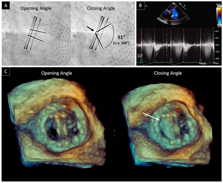
Figure 1.
Bileaflet mechanical mitral valve obstruction. (A) Fluoroscopic evaluation of normal opening and abnormal closing angles in a patient with prosthetic mitral valve dysfunction. The dotted line refers to the normal leaflet closure. The abnormal leaflet closure was confirmed by the lack of leaflets contact at the hinge area (black arrow). (B) Holosystolic regurgitation at continuous wave Doppler by TEE. (C) 3D view of prosthetic mitral valve from atrial side. During systole, only one leaflet closes (white arrow).

