Figure 2.
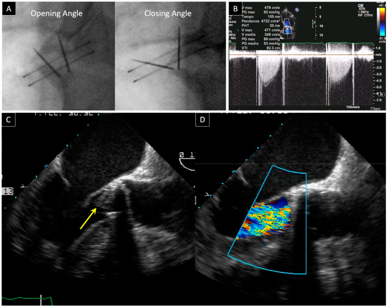
Bileaflet mechanical aortic valve obstruction. (A) Fluoroscopy shows normal opening and abnormal closing angles in a patient with prosthetic aortic valve dysfunction. (B) Flow acceleration of the anterograde flow is identified with color flow imaging from the TTE apical approach and it is associated with high transprosthetic gradients at continuous wave Doppler (ΔPmean 53 mmHg). (C) 2D TEE from a 120° view reveals a thrombotic hyperechogenic mass on the ventricular side of the prosthesis (arrow). (D) Severe intraprosthetic regurgitation as assessed by 2D TEE.

