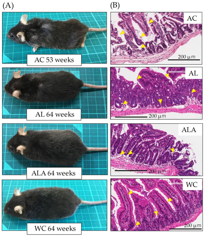
Figure 3.
Observations of the appearance (A), small intestine tissues (B), in mice at 53 weeks of age (AC) or 64 weeks of age (AL, ALA, and WC). (B) Histological observations by H&E staining of small intestine tissues. Arrows indicate the destruction of the jejunal epithelium. Original magnification: 200×. Scale bar = 200 μm. AC, AL, or ALA mice were fed a normal diet, containing limonoids alone, and L-arginine + limonoids, respectively. WT mice (WC) were fed a normal diet.

