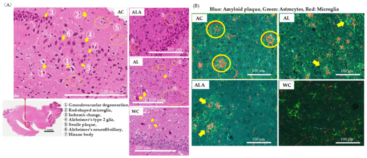
Figure 6.
Histopathological analysis of brain tissues of 53-week-old (AC) or 64-week-old (AL, ALA, and WC) mice. (A) H&E staining of the regions around the hippocampus (original magnification: 200×, scale bar = 200 μm) and (B) immune triple fluorescence staining of regions around the hippocampus (original magnification: 400×, scale bar = 100 μm) of AC, ALA, AL, and WC mice. The sections show senile plaques formed with the amyloid β storage cells. (B) Triple immunofluorescence staining images of hippocampal samples localized Amylo-Glo-stained amyloid plaques (blue), GFAP-positive astrocytes (green), and IbA 1-positive microglia (red) on the same section. In the AC mice, hypertrophied astrocytes and activated microglia were upregulated around the amyloid plaques, forming senile plaques.

