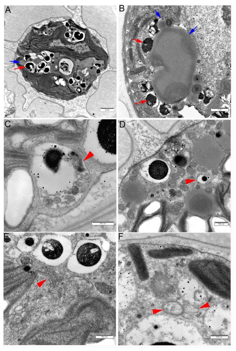
Figure 6.
Ultrastructure of cells of Haematococcus lacustris cultures after three days of exposure to 0.8% NaCl. (A) A whole cell; red arrow points at an osmiophilic granule probably containing carotenoids including astaxanthin; blue arrow points at a lipid droplet. (B) Part of a cell showing lipid droplets (blue arrows) with ‘carotenoid bodies’ docking on them (red arrows). Osmiophilic ‘carotenoid bodies’ are located within single-membrane small vacuoles probably representing autolysosomes. (C) An autolysosome containing thylakoid remnants (red arrowhead). (D) Part of a cell showing electron-translucent lipid droplets, an electron-dense osmiophilic ‘carotenoid body’ within an autolysosome, and a small ‘carotenoid body’ surrounded by a double-membrane autophagosome (red arrowhead). (E) Several ‘carotenoid bodies’ in the vicinity of Golgi cisternae (red arrowhead). (F) Autophagosome (left red arrowhead) and a structure probably representing an omegasome with an expanding phagophore (right red arrowhead).

