
Fig. 1.
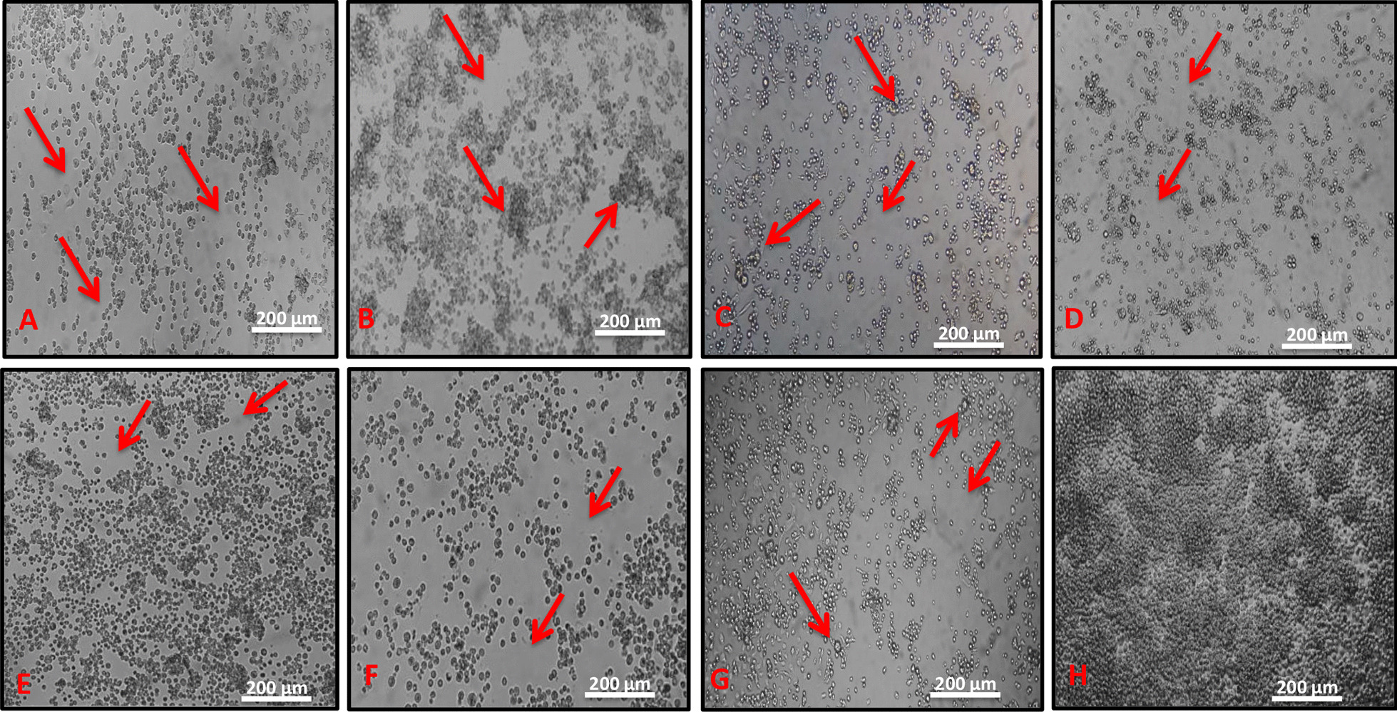
Cytopathic effect (CPE) observed in infected C6/36 cells (red arrow). a Cells inoculated with the sample BE AR 805503 (CPE at 6th day post-infection), showing destruction of cell’s monolayer; b cells inoculated with the sample BE AR 805529 (CPE at 4th day post-infection), showing destruction of cell’s monolayer and formation of clumps; c cells inoculated with the sample BE AR 805511 (CPE at 6th day post-infection), showing destruction of cell’s monolayer and large cells (larger than normal cells); d–f cells inoculated with the sample BE AR 805514 (CPE at 6th day post-infection), BE AR 820396 (CPE at 6th dpi) and BE AR 805520 (CPE at 6th day post-infection), respectively, showing destruction of the cell’s monolayer; g cells inoculated with the sample BE AR 805525 (CPE at 6th day post-infection), highlighting destruction of the cell’s monolayer and large cells (larger than normal cells); h negative control (C6/36 cells) showing no CPE (100X)