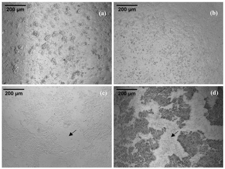
Figure 4.
Madin–Darby canine kidney II (MDCK II) cell culture after application of 100 µM Checacin1 and its truncated forms. (a,b): intact cell layer after application of Checacin11−11 and Checacin112−25 (c,d): disintegrated cell layer after application of Checacin11−21 and Checacin1. Arrows indicate areas of disintegrated cell layers.

