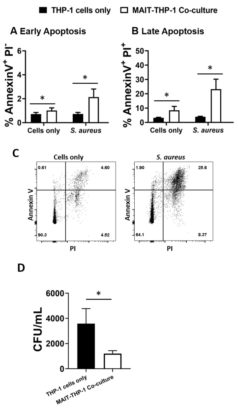
Figure 5.
MAIT cells prevent S. aureus intracellular survival within THP-1 cells. THP-1 cells (5 × 10⁵/mL) were infected with S. aureus (strain PS80) at MOI 1 for 3 h before elimination of extracellular bacteria by gentamicin treatment. THP-1 cells were then either cultured alone or co-cultured with uninfected MAIT cells (5 × 10⁵/mL) for 24 h, after which cells were either collected for analysis of apoptosis by Annexin V and PI staining (A–C) or washed in PBS and lysed in 0.1% Triton X-100 for 10 min (D). Lysates were plated on TSA plates and incubated overnight. CFUs were enumerated the following day, and CFU/mL of original wells calculated. Results are expressed as mean percentage of apoptotic cells + SEM (A,B) or mean CFU/mL + SEM (D). Representative FACS plots are shown (C). n = 5–6 MAIT cell donors per group. ‘Cells only’ refers to uninfected cultures. Statistical analysis by pairwise Wilcoxon signed rank. * p ≤ 0.05.

