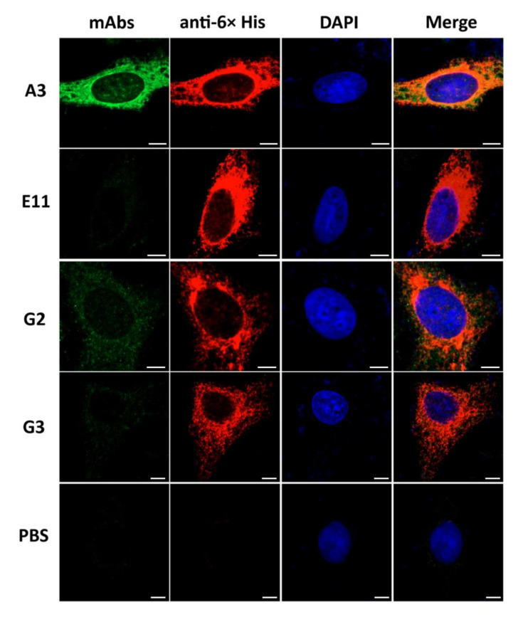
Figure 3.
Co-localization of the rS1-specific-mAbs to PEDV S1 overexpressed in HeLa cells as determined by confocal microscopy. HeLa cells transfected with S1-pTriEx 1.1 recombinant plasmid were cultured on cover slips in 24-well culture plate for 48 h. Cells were fixed with cold acetone-methanol (1:1), followed by staining with 10 µg of individual mAbs to S1 (anti-rS1) and rabbit anti-His antibody (1:500) as primary antibodies and fluorophore-conjugated secondary antibodies (1:300). mAbA3, mAbG2 and mAbG3 (green) colocalized with intracellular S1 overexpressed in the HeLa cells (red) and seen as orange or yellow matter in merge. The mAbE11 did not bind (or bound negligibly) to the overexpressed S1 in the transfected cells. Nuclei stained blue by DAPI dye. Scale bar = 5 µm.

