Figure 1.
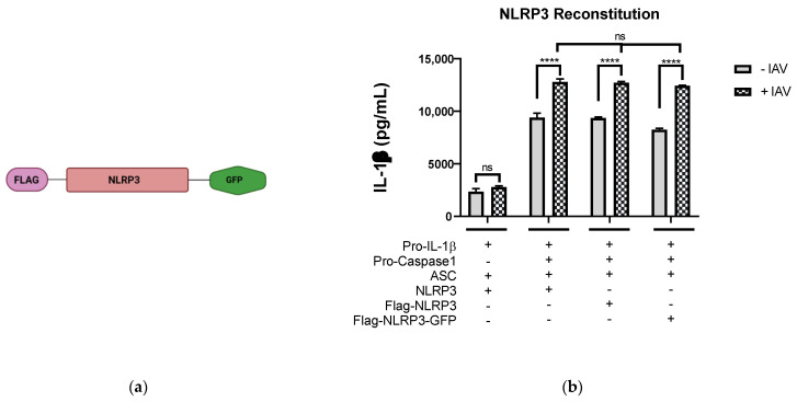
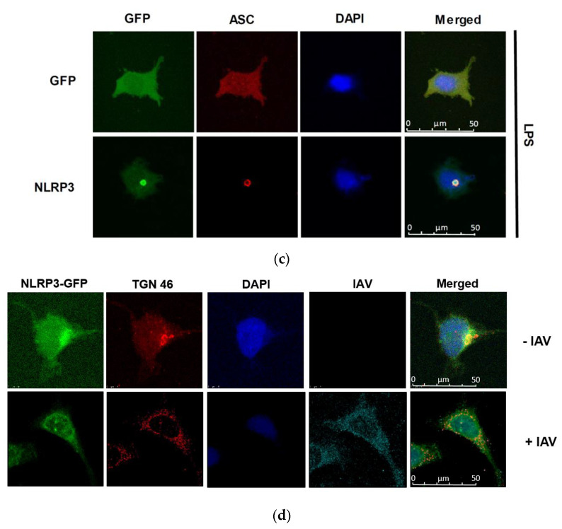
IAV infection induces the formation of dispersed trans-Golgi network. (a) Schematic representation of Flag-NLRP3-GFP construct. (b) HEK293T cells were transfected with different combinations of plasmids expressing porcine NLRP3, ASC, pro-caspase-1, and pro-IL-1β as indicated. At 12 h.p.t., the cells were mock-infected or infected with Sk02 at an MOI of 10. Porcine IL-1β from the cell-free supernatants at 12 h.p.i was measured by ELISA (two-way ANOVA; **** p < 0.0001; ns- Not significant). (c) HeLa cells were co-transfected with ASC together with either GFP or NLRP3-GFP and were stimulated with LPS (200 ng/mL) for 80 min. The cells were fixed, permeabilized, blocked, and probed with appropriate antibodies, followed by DAPI staining. GFP/NLRP3-GFP (green), ASC (red), and nucleus (blue) were visualized by confocal microscopy. Scale bar, 50 μm. (d) HeLa cells expressing NLRP3-GFP were infected with Sk02 at an MOI of 10. At 80 m.p.i, the cells were subjected to immunofluorescence for NLRP3-GFP (green), TGN (red), IAV (cyan), and nucleus (blue). Scale bar, 50 μm.


