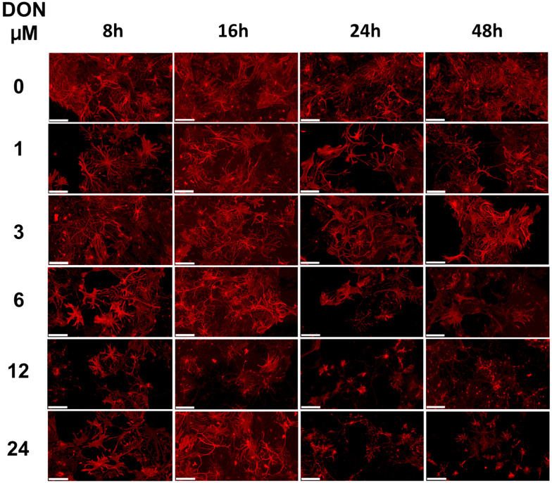
Figure 6.
Impact of DON exposure on glial cells in primary hippocampal cultures. Three independent cultures were prepared and analysed for changes in the astrocyte morphology using GFAP staining. Representative images upon treatment with 0, 1, 3, 6, 12, or 24 µM DON for 8, 16, 24, or 48 h. Scale bars: 100 µm.

