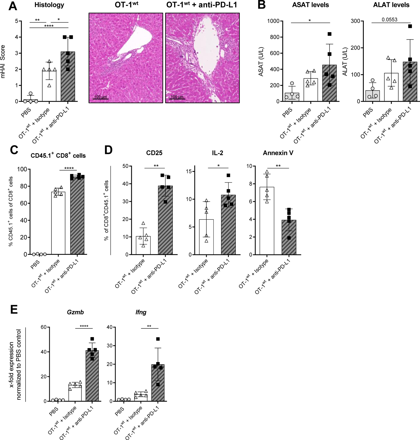
Fig. 5. Inhibition of PD-L1 induces aggravated cholangitis in K14-OVAp recipient animals.

Cholangitis severity was assessed 5 days after adoptive transfer of OT-1wt CD8+ T cells into K14-OVAp mice treated with anti-PD-L1 or isotype-control. (A) mHAI histological activity index of H&E stained liver sections and (B) serum levels of liver transaminases ASAT and ALAT. (C) The percentage of transferred OT-1wt CD8+ CD45.1+ T cells was determined within recipient livers and (D) the expression of CD25, IL-2 and annexin V on OT-1wt CD8+ T cells isolated from livers using flow cytometry. (E) Expression of Gzmb, Ifng and Pdcd1 mRNA was analyzed in K14-OVAp whole liver tissue by qPCR. Data are expressed as mean ± SD; n=4–5, Data from one representative experiment of two independent experiments is shown. Levels of significance: *p≤ 0,05; **p≤ 0,01; ****p≤0,0001 (ordinary one-way ANOVA (A, B, C, E), Mann-Whitney U test (D)).
