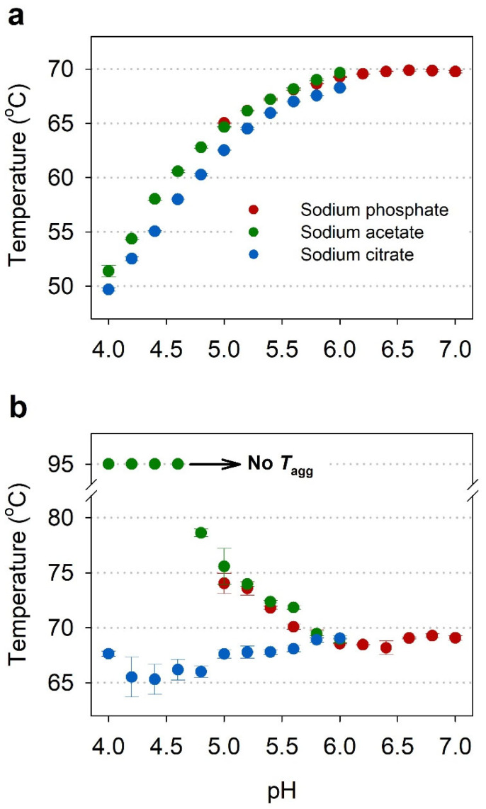
Figure 3.
Effect of pH and buffer species on Tm (a) and Tagg (b) of IgG in three different buffers (red—sodium phosphate; green—sodium acetate; blue—sodium citrate), as measured by nanoDSF. (a) Graph of Tm of IgG as a function of pH; (b) Graph of Tagg of IgG as a function of pH. All samples were set at a concentration of 1 mg/mL and values are the mean and standard deviation of triplicate measurements.

