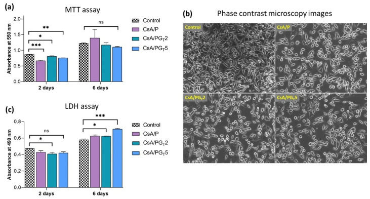
Figure 8.
In vitro biological analysis of nanofibrous membranes. (a) MTT assay—viability and proliferation of NCTC fibroblast cells grown onto the surface of nanofibrous membranes, after 2 and 6 days (ns p > 0.5, * p < 0.05, ** p < 0.005, *** p < 0.0005). (b) Phase contrast microscopy images of NCTC cells. (c) LDH assay—quantification of dead NCTC fibroblasts after 2 and 6 days (ns p < 0.5, * p < 0.05, *** p < 0.0005).

