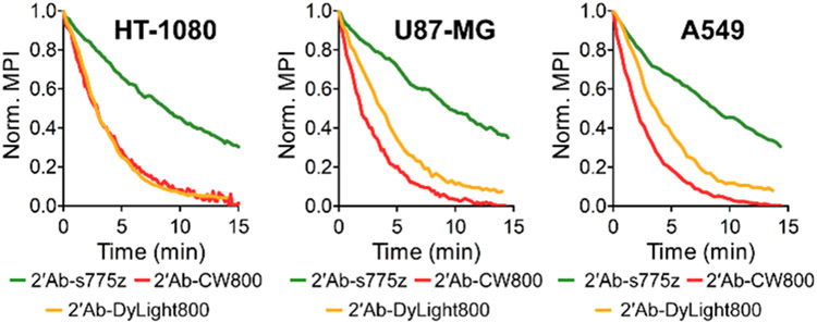
Figure 2.
Photostability comparison of secondary goat anti-rabbit IgG antibodies labeled with NIR dyes. Cells were incubated with primary anti-Tubulin and fluorescent secondary antibody (2’Ab-s775z, 2’Ab-CW800, or 2’Ab-DyLight800) followed by continuous irradiation with NIR light within a microscope equipped with an ICG filter (ex: 769/41, em: 832/37). Plots show normalized mean pixel intensity (MPI) for time-lapse cell micrographs.

