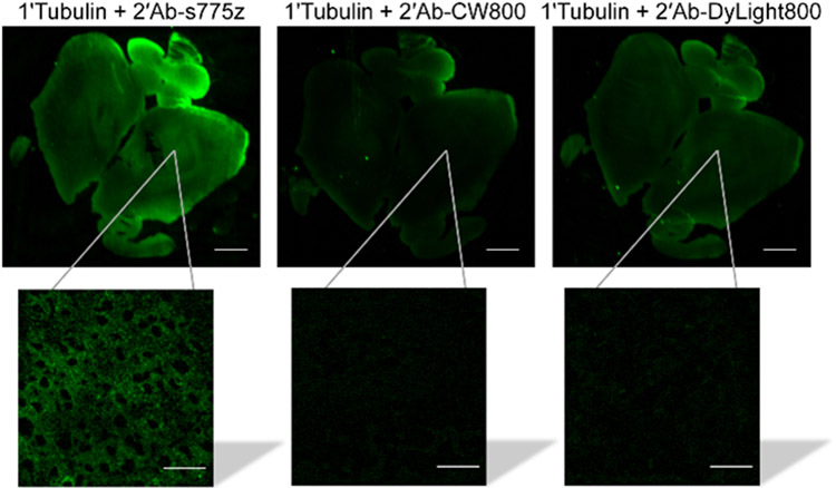
Figure 4.
Comparison of secondary goat anti-rabbit IgG antibodies labeled with NIR dyes for targeting primary anti-Tubulin in mouse brain tissue. Mouse brain tissue slice was incubated with anti-Tubulin overnight at 4 °C and incubated the following day with a fluorescent secondary antibody (2’Ab-s775z, 2’Ab-CW800, or 2’Ab-DyLight800) for 2 hr at room temperature. Each NIR fluorescent image is a brain tissue slice with the inset showing a magnified micrograph. Scale bars on the brain tissue and micrograph inset are 1 mm and 30 μm, respectively.

