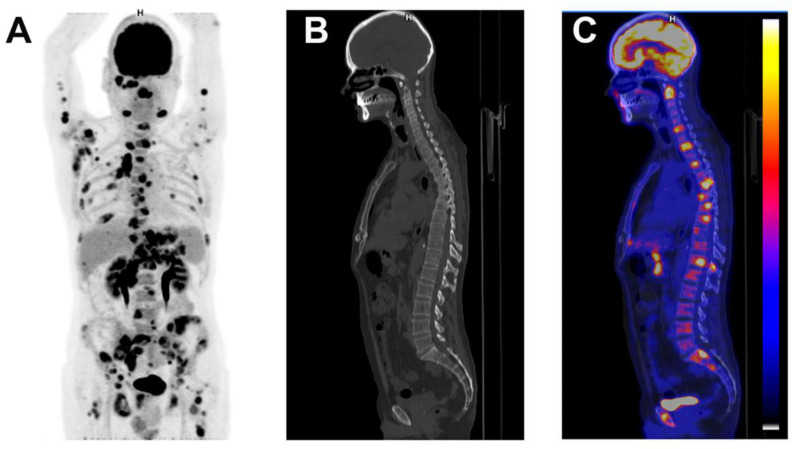
Figure 4.
Patient with diffuse large B cell lymphoma. A 52-year-old man was admitted with 7 week’s duration of FUO, sweating, myalgia and lower back pain. Physical examination showed axillary lymph nodes. Abdominal examination did not show hepatosplenomegaly. Laboratory testing showed a CRP of 132 mg/L with normal leucocytes. ASAT was 43 U/L, ALAT 67 U/L, alkaline phosphatase 155 U/L and. Angiotensin converting enzyme was normal. Blood cultures were negative as were bartonella, Q Fever, mycoplasma, chlamydia, HVC, HBV and toxoplasmosis serologies with past EBV and CMV immunity. The CAPCT scan performed 2 weeks before hospitalization was normal. Coronal maximum intensity projection FDG-PET (A), sagittal low-dose CT (B) and fused FDG-PET/CT (C) demonstrated intensive FDG uptake in axillary, submandibular, mediastinal, para-aortic, epigastric and inguinal lymph nodes and in bones. A submandibular lymph node confirmed the diagnosis of diffuse large B cell lymphoma.

